Modern medical advancements have significantly improved the diagnosis and treatment of various conditions, particularly those involving the digestive system. Among these techniques, endoscopy has become a cornerstone of investigation, offering a minimally invasive way to visualize internal structures and collect tissue samples for further analysis. This article delves into the intricate relationship between endoscopy and biopsy, explaining how tissue samples are collected and why they are crucial for accurate diagnoses.
What is Endoscopy?
Endoscopy is a medical procedure used to examine the interior surfaces of organs and tissues within the body. It involves the use of an endoscope, a long, flexible tube equipped with a light source and a camera. This device allows healthcare professionals to capture real-time images of the area under examination, such as the esophagus, stomach, intestines, or lungs.
Endoscopy is performed for both diagnostic and therapeutic purposes. It can identify abnormalities like ulcers, inflammation, and tumors while simultaneously enabling interventions, such as removing polyps or stopping internal bleeding. This dual functionality makes it an indispensable tool in modern medicine.
What is a Biopsy?
A biopsy is a procedure in which a small tissue sample is collected from the body for microscopic examination. This process allows pathologists to evaluate the cellular structure of the sample, detecting abnormalities that may not be visible through imaging alone.
Biopsies are essential for diagnosing a range of conditions, including infections, inflammatory diseases, and cancers. By providing detailed insights into cellular changes, a biopsy often forms the basis for accurate treatment planning.
The Role of Endoscopy in Biopsy
Endoscopy is often paired with biopsy procedures to enhance diagnostic accuracy. The ability of an endoscope to provide high-resolution visuals of internal structures enables targeted tissue collection. This precision is particularly important when investigating small lesions, areas of inflammation, or suspicious growths.
For example, during a gastroscopy (a type of endoscopy used to examine the stomach and upper digestive tract), a doctor can identify areas that appear abnormal and collect tissue samples using specialized instruments attached to the endoscope. Similarly, in a colonoscopy (an endoscopic examination of the colon), polyps can be removed and analyzed to rule out malignancy.
How Tissue Samples Are Collected During Endoscopy
The process of collecting tissue samples during endoscopy involves several meticulous steps. Here’s how it works:
1. Preparation for the Procedure
Before the endoscopy, patients are typically advised to follow certain dietary restrictions and may be asked to fast for several hours. For endoscopies of the digestive tract, bowel-cleansing preparations may be necessary to ensure a clear view. Sedation is often administered to keep the patient comfortable throughout the procedure.
2. Visualization of the Area
Once the endoscope is inserted into the target area, the doctor carefully examines the internal surfaces. Advanced endoscopic equipment often includes features like magnification and narrow-band imaging, which help identify subtle abnormalities that may require further investigation.
3. Collecting Tissue Samples
When an area of interest is identified, the doctor uses special biopsy forceps or needles attached to the endoscope to extract a small tissue sample. This step is carefully performed to ensure that the sample is representative of the abnormality while minimizing damage to surrounding tissues.
For certain types of endoscopies, such as bronchoscopy (used to examine the lungs), a needle biopsy may be performed to collect tissue from deeper layers.
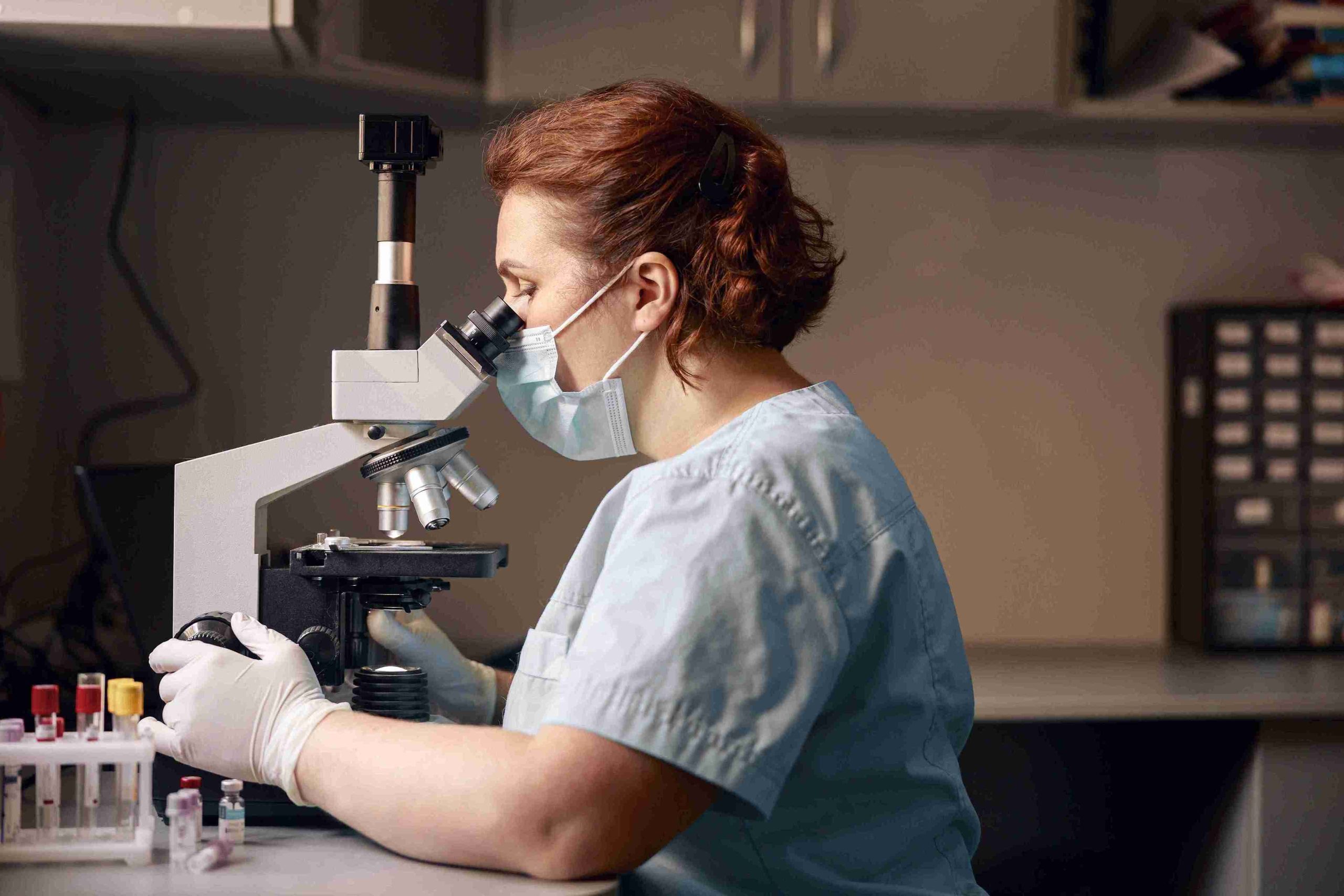
4. Handling the Tissue Samples
Once collected, the tissue samples are carefully placed in containers with preservative solutions to maintain their integrity. These samples are then sent to a pathology lab for analysis.
The Importance of Biopsy in Diagnosing Diseases
While endoscopy provides visual evidence of abnormalities, a biopsy is crucial for confirming the nature of those abnormalities. This combination is particularly valuable in diagnosing conditions such as:
- Cancer: A biopsy can differentiate between benign and malignant growths, providing essential information about the type and stage of cancer.
- Infections: Tissue samples can reveal the presence of bacteria, viruses, or fungi, enabling targeted treatment.
- Inflammatory Conditions: Conditions like Crohn’s disease or gastritis can be confirmed by analyzing cellular changes in the tissue.
Without a biopsy, visual observations alone may lead to inconclusive or incomplete diagnoses, potentially delaying appropriate treatment.
Advances in Endoscopic Biopsy Techniques
Technological advancements have revolutionized the way tissue samples are collected during endoscopy. Innovations such as endoscopic ultrasound (EUS) and confocal laser endomicroscopy have enhanced the precision and effectiveness of biopsies.
- Endoscopic Ultrasound (EUS): EUS combines endoscopy with ultrasound imaging, allowing for detailed visualization of deeper layers of tissue. This technique is particularly useful for diagnosing conditions affecting the pancreas, liver, and lymph nodes.
- Confocal Laser Endomicroscopy: This cutting-edge technology provides real-time microscopic views of tissue, enabling immediate evaluation of cellular structures without the need for external lab analysis.
These advancements continue to push the boundaries of what endoscopy can achieve, reducing the need for more invasive procedures.
Risks and Complications of Endoscopic Biopsy
Although endoscopy and biopsy are generally safe procedures, they are not without risks. Potential complications include bleeding, infection, and perforation of the organ being examined. However, these risks are rare and are typically outweighed by the benefits of accurate diagnosis and timely treatment.
Patients are encouraged to discuss their medical history and any concerns with their doctor before undergoing the procedure. Proper preparation and adherence to post-procedure care instructions can further minimize risks.
The Future of Endoscopy and Biopsy
The field of endoscopy continues to evolve, with ongoing research focusing on enhancing diagnostic accuracy and patient comfort. Developments such as artificial intelligence (AI)-assisted endoscopy are being explored to improve the detection of subtle abnormalities.
Furthermore, minimally invasive techniques are becoming more refined, reducing recovery times and allowing for earlier interventions. These advancements hold promise for even more effective diagnosis and treatment of conditions affecting internal organs.
Conclusion
Endoscopy and biopsy are complementary procedures that play a vital role in modern medicine. While endoscopy provides a detailed visual assessment of internal structures, the biopsy offers microscopic insights that are critical for accurate diagnosis. Together, they enable healthcare professionals to detect, analyze, and address a wide range of conditions, from infections to cancers.
As medical technology continues to advance, the capabilities of endoscopic biopsy are expected to expand further, paving the way for more precise and effective healthcare solutions. For patients experiencing unexplained symptoms or abnormalities, timely endoscopy and biopsy can make all the difference in achieving a positive outcome.